Posts

Combating Digital Vision Syndrome

Digital Vision Syndrome has been a popular topic in the last few years as more of our time is dedicated to viewing our many digital devices.  After long periods of use, many individuals, including myself, experience discomfort and visual problems.  

What kind of symptoms are we talking about? Eyestrain, headaches, and dry eyes to name a few of the major offenders.   The causes of Digital Vision Syndrome, however, are a little more difficult to pinpoint.  It is typically a combination of various factors such as: poor lighting, posture, blue light emitted, uncorrected visual problems.  

We take several steps to combat Digital Vision syndrome. Our first line of defense focuses on how we view our devices.  Basic ergonomics such as positioning yourself at a computer screen so that the screen is 20 to 28 inches away from your eyes and between 15 to 20 degrees below eye level. Additionally, proper room lighting is imperative.  Also make sure to take breaks by spending at least 20 seconds to look at something 20 feet away every 20 minutes of computer use.  This is called the 20-20-20 rule.  

The second line of defense focuses on blocking the harmful blue light emitted from these devices. Research has shown that certain blue light has been linked to sleep issues and has a possible link to certain eye diseases.  Special lens coatings can be added to your glasses to reduce this light. 

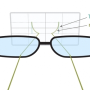
The third line of defense focuses on reducing the amount of eyestrain by reducing the amount of focusing work your eyes do when looking something near and the amount of tension on the eye muscles.  The newest technology in this category has been the Neurolens® released by eyeBrain Medical.  Using the Neurolens Measurement Device, eye alignment is accurately measured for all distances.  Any eye misalignment can cause the visual system to work harder which can lead individuals to experience the symptoms of Digital Vision Syndrome.  The Neurolens® is a special lens design that corrects eye misalignment. By reducing the demand on the visual system symptoms of Digital Vision Syndrome can be greatly reduced or eliminated.  

Blog contribution by Jonathan Kiriboon, Optometry Intern, College of Optometry, Western University of Health Sciences.

Sources:

Scleral Contact Lenses: Are They For You?

Scleral contact lenses are large, gas permeable lenses that rest on the white part of the eye known as the sclera. This differs from soft contact lenses which we fit to the cornea, the dome-shaped front of the eye. This unique design allows the lens to functionally replace the irregular cornea with a smooth optical surface to correct vision problems such as keratoconus (thinning and conical protrusion of the cornea) and other corneal irregularities. The space between the cornea and the back surface of the scleral lens acts as a liquid reservoir. This reservoir is filled with tears and provides comfort by lubricating the surface of the cornea to reduce dry eyes. 

Practically a scleral lens is a prosthetic cornea that recreates a perfect optical surface and makes vision a whole lot better. Let’s look at the following images that show what these menses look like:

pastedGraphic.png
pastedGraphic_1.png

Scleral contact lenses are custom-made for patients and can provide better comfort and vision for those who have severe forms of:

• Keratoconus

• Ocular surface diseases (Sjogren’s, Steven’s Johnson Syndrome)

• Irregular astigmatism

• Highly irregular corneal transplants

• Post-refractive surgery complications (LASIK, PRK)

• Dry eye

Previously, scleral lenses have been reserved for only complex corneal and contact lens cases but are now gaining popularity and are being used for normal patients. Its distinct design provides increased comfort and stability on the eye leading to consistent vision, reduced glare and reduced dryness. Patients that have experienced poor comfort or vision with regular contacts may be good candidates for scleral lenses. 

Sources:

http://www.reviewofcontactlenses.com/content/d/rgp_lenses/c/52262/
http://www.allaboutvision.com/contacts/scleral-lenses.htm
http://cdna.allaboutvision.com/i/contacts-2016/scleral-lens-diagram-330×[email protected]
http://cdna.allaboutvision.com/i/contacts-2016/scleral-lens-in-eye-330×[email protected]

Neurolenses

Eye Motor Deficiencies:  Teaming, Focusing and Muscle Deficiencies

Clear vision is not the only important factor for comfortable vision. Equally important is when your eyes efficiently work together. This includes proper eye alignment and the ability to move the eyes smoothly together in all directions. Additionally, the tiny muscles located inside the eye must efficiently control our ”zoom” ability or near focus. Our brain, specifically our trigeminal nerve, senses when our eyes are out of alignment and compensates to help improve our focus. All of the compensation that we do can cause symptoms such as fatigue, headaches, neck pain, and discomfort in the eyes (asthenopia).

A variety of methods are used to help correct these vision-related issues. These include the standard methods of glasses or contact lenses but may also include Neurolens, prisms or over/under correcting lenses. Prisms can be prescribed in glasses that bend light and allow for comfortable vision when your eyes are not aligned normally. Over or under- correcting lenses serve to relieve or stimulate accommodation (focusing).

In the last few years we have improved our treatment of many eye misalignments. Using what is called a neurolens, we are able to better correct eye teaming issues. What is special about neurolens is its ability to correct distance ad near deviations in a single lens. This allows patients to be optimally corrected full-time. In fact, many patients respond so well that their symptoms are greatly reduced or eliminated after using the lenses for only a few weeks. If we are talking about a migraine or significant headache this can be life changing!

Yoga and Glaucoma (Say that 5 times fast)

Glaucoma is one of the leading causes of irreversible blindness because of damage to the optic nerve when fluid pressure increases in the eye. Doctors advise patients to live healthy and active lifestyles, but there are certain activities that should be avoided by glaucoma patients like pushups and lifting heavy weights due to elevated intraocular pressure (IOP). A new study has shown the potential risk associated with various yoga positions and exercises that involve inverted poses. In this research, both normal and glaucoma patients did four yoga positions. The greatest increase in pressure was found with the downward facing dog position where you place both feet and hands on the floor while looking at the ground. “The measurements were taken after the participants retuned to a seated position and again after waiting ten minutes, the pressure in most cases remained slightly elevated from the baseline.” In previous studies, participants in the headstand position showed almost two-fold rise in IOP. It is now advised to inform and educate glaucoma patients about the risks of physical exercises like yoga, so progression of glaucoma doesn’t get worse. Many times we consider a point or two of pressure increase to be a reason to prescribe additional eye drops or to recommend a glaucoma surgery. When patients go to yoga, patients should tell their instructors their disease to modify their yoga practice.

Mount Sinai Health System. (2016, January 7). Certain yoga positions may impact eye pressure in glaucoma patients. ScienceDaily. Retrieved January 20, 2016 from www.sciencedaily.com/releases/2016/01/160107105234.htm

Makeup, Keep it Clean!

A light application of makeup can often help you to look and feel your best. Without proper care, however, it may impair your capacity to see your best.  Whether you are going for a natural look or something more dramatic, it is important to use discretion when applying around your eyes.

Keep it clean:                                   

Make sure your hands are washed before you begin.  Use only your own makeup and brushes.  It may seem fine to borrow a swipe of mascara, but even your BFF may have germs that are harmful to you.  Infections from poor hygiene or contamination may lead to itchy, swollen eyes and light sensitivity.

Do not disturb:                                   

Only apply eye makeup in a setting where you can be relaxed and focused.  Putting on makeup in a vehicle risks injury at every stop and bump during your commute.  Take your time and don’t rush; not only will you get better results, but you’ll do better to avoid poking yourself in the eye or scratching your cornea.

Contact lens wearers:                 

It is better to put in your soft contact lenses before your makeup routine.  When choosing a mascara, avoid mascara with lengthening fibers, which can scratch, and waterproof mascara, which may stain your lenses.  At the end of the day, remove your contacts, clean them, and place in fresh solution before you wash off your makeup.

It can be rewarding to use cosmetics, but safety should always be top priority.  If your makeup causes you to experience irritation or injury, discontinue use and contact your optometrist.


Blog contribution by Kaitlyn Sanchez, Optometry Intern, College of Optometry, Western University of Health Sciences.


http://www.aoa.org/patients-and-public/eye-and-vision-problems/glossary-of-eye-and-vision-conditions/conjunctivitis?sso=y

http://www.aoa.org/patients-and-public/caring-for-your-vision/contact-lenses/contact-lenses-and-cosmetics?sso=y

http://www.fda.gov/Cosmetics/ProductsIngredients/Products/ucm137241.htm

Healing the Eye

Our eyes are exposed to a wide variety of environmental conditions such as extreme weather, work environment and wearing contact lenses.  Sometimes these conditions may be so harsh that they stress the cornea, the outermost eye structure. Occasionally a patient will present in my office with an extremely compromised cornea. Such cases sometimes need a form of an eye band-aide to properly heal.

Enter the Amniotic Membrane

Such a bandage is an amniotic membrane which is derived from the placenta of an elective c-section. Human amniotic membrane is a unique collagenous membrane derived from the innermost submucosa of the placenta. Harvested under sterile conditions from the placenta of elective C-section after a full term pregnancy in medically cleared donors. This type of tissue has been used widely in the treatment of surface ocular diseases as it aids epitheliazation, reduces inflammation and fibrosis, prevents structural damage, and is also known to have some antimicrobial properties. It is used for a wide variety of ocular surface disorders such as: corneal ulcers, chemical or thermal burns of the cornea, and persistent epithelial defects.

An example used in our office is the ProKera Ring. It is a cryopreserved, sutureless amniotic membrane clipped to a plastic ring. ProKera is placed on the eye in a similar fashion as a large contact lens. ProKera does not require an operating room and can be performed in the office. Most importantly, it is reasonably well tolerated by patients.

Blog contribution by Lilia Babakhan, Optometry Intern, College of Optometry, Western University of Health Sciences.


The Future of Glasses?

In recent years, there have been innovations in eyewear aimed at helping those suffering from presbyopia, the gradual inability of the eyes to focus due to age. Presbyopia usually begins starting at around age 40 and can affect anyone, even if you have never worn glasses before. According to the World Health Organization, more than one billion people in the world were presbyopic as of 2005. Current eyewear treatments include bifocal and Progressive lenses, but advancements in technology offer new solutions.

One example is Superfocus, which is based in California. These glasses have an adjustable slider on the bridge that lets you change the prescription from distance to near or anywhere in between. This is accomplished by a pocket of soft silicone between two lenses that changes the shape of one of the lenses, and therefore the prescription. This mimics the movements of the lens in the pre-presbyopic eye. The other lens is removable and can be switched to a tinted lens, for example.

Another lens innovation was called emPower, a line of electronic eyeglasses offered by Virginia-based company, PixelOptics from 2011 to 2013. These glasses allowed the wearer to switch between two prescriptions, near and distance. They could be programmed to react either to a fingertip pressing the temple of the frame or to a downward tilt of the head, which triggered an accelerometer. The lenses consisted of LCD materials between two layers of plastic and when an electrical charge hit the LCD, the molecules realigned and the prescription changed. The near prescription was made up of an oval-shaped area in the bottom half of the lenses that disappeared instantaneously when the distance prescription was triggered. In terms of price, the glasses retailed from $1,000-$1,500. Besides being a costly investment, there were issues with emPower’s optics, electronics and design, and PixelOptics declared bankruptcy in November 2013. Although these particular glasses are no longer being offered, the technology exists and perhaps with some modifications, a similar concept could revolutionize the presbyopic eyewear industry.

We’ll keep our eyes open and report on it here at the BOG Blog when a new prospect arises!

Blog contribution by Lorraine Lopez, Optometry Intern, College of Optometry ,Western University of Health Sciences