
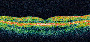
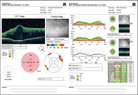
Optical Coherence Tomography allows us to scan under the retina and optic nerve with a laser. This is a very important instrument that gives us much more information than a digital image, as it allows us to measure swelling or detachments under the retina. Additionally, we can follow glaucomatous tissue changes in the optic nerve. It will take three minutes.
How does it work?
You look into the instrument and fixate on a light. Dr. Bender will obtain a nice color map illustrating thickness and values of the optic nerve or central retina, together with side profiles of the tissue. He can determine if there is swelling in the retina due to macular degeneration or diabetes and he can track glaucoma progression.