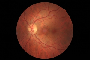
When we need to get a closer look at conditions of the retina and the optic nerve we use Retinal Imaging. In this examination, the digital camera takes high resolution images of the inside structure of the eye, the retina. This examination takes only a couple of minutes.
Retinal Imaging is most commonly used to examine glaucoma, but it could also be used for growths such as retinal freckles or diabetic retinopathy.
How does it work?
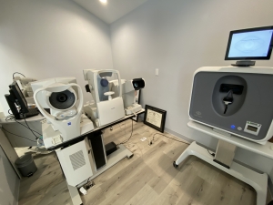
You place your forehead and chin in a headrest and stare at a point of a light target. Then you will see a bright flash like from a camera.
The technician will view your eye in a monitor. When Dr. Bender receives the image, he will review it with you. If you have done this before he can compare it with the earlier images and it is easy to see if there are any changes.
The image of your eye can be analyzed using various color filters and zoom features – it is quite fascinating to look at!
We offer Retinal Imaging as an optional yearly screening test for everyone who wants to monitor the health of their eyes. The cost is $39.00.
Please note
In some cases we need to dilate the pupil for this examination, please read more under Dilate pupils.


